Sunday 31 March 2019

Stelar Evolution in Pteridophytes


Write as second assignment

Stelar Evolution in Pteridophytes

Central cylinder or core of vascular tissue consisting of xylem, phloem, pericycle and sometimes medullary rays and pith is called Stele.
The word stele is derived from Greek which means  central pillar. The Stele is the central cylinder and is separated from the cortex by the endodermis.  The endodermis is the innermost layer of the cortex and pericycle is the outermost portion of the Stele.
Van Tiegam and Douliot proposed Stellar Theory in 1886.
According to to the the stellar theory there is no fundamental difference in the gross anatomy of the stem and root because in both of them is Stele surrounded by cortex is present
According to this theory the stele is not only made up of xylem and phloem but the tissue like pericycle, vascular rays and pith are also associated with it.

Types of steles
Easu and Smith recognise two principal types of stelar organisation among the vascular plants.  These are the Protostele and Siphonostele.
Protostele
The name protostele was suggested by Jeffrey and he regarded it as the most primitive and simplest type of stele.
Steel in which the gas cylinder consists of a solid core of xylem surrounded by phloem pericycle and endodermis is called protostele. There is no pith in a protostele. it is a fundamental type of Steel of vascular plants from which the other type originated in the course of evolution.
Brenner classified the protostele into following types
Haplostele: Protostele with smooth core of  xylem surrounded by uniform layer of phloem is called haplostele.  It is found in several fossil genera like Rynia and Horneophyton and living genera like Selaginella kraussiana.

Actinostele: a protostele in which xylem core is stellate or star shaped with radiating arms is called at actinostele. actor no still the phloem is not present in a continuous manner but in the form of separate groups which alternate with the arms of the star shaped xylem. Example Psilotum,  Lycopodium serratum.

Sometimes the actinostele may show variations due to breaking of xylem tissue in two different forms. The common modifications of actinostele are
Plectostele: The xylem gets broken into a number of more or less parallel plates and such xylem plates alternate with phloem plates.  Example Lycopodium clavatum and Lycopodium volubile.

Mixed protostele in this type of stele xylem appears in the form of irregular crops that are scared scattered in the ground mass of the phloem. E.g., L. cernuum.


Siphonostele : It is a modified form of protostele in which the pith is present in the centre of the stele. A medulated protostele  is called as siphonostele. During the development of siphonostele core of xylem is replaced by parenchymatous pith.
In siphonostele the central pith is surrounded by a cylinder of xylem and phloem. This type of stele is found in members of Pteropsida.
Origin of Siphonostele:
There is a general acceptance that the Siphonostele has evolved from protostele. Two theories have been proposed for evolution of Siphonostele from protostele,
I Extrastelar origin of pith: This theory was put forwarded by Jeffrey. According to him the pith originated as a result of the invasion of cortical parenchymatous cells into the stele. This invasion occurs through the leaf gaps and branch gaps.
II Intrastelar origin of pith:
Boodle, Gwynne-Vaughan, Bower supported this theory of intrastelar origin of pith.
According to this theory the innermost or centrally placed vascular tissue changes into parenchymatous cells which behave as pith. Presence of isolated trachieds in the pith in Botrychium viriginianum, Osmunda regalis support the intrastelar origin of pith.

Types of Siphonostele

Jeffrey classified the siphonostele into following two types on the basis of the position of phloem :
Ectophloic Siphonostele:
in this the earth is surrounded by xylem which in turn is surrounded by phloem, pericycle and endodermis.  So in this case phloem is present only external to the xylem. E.g., Osmunda


Amphiphloic Siphonostele:
In this case the phloem is present on both the sides of xylem i.e., on external as well as internal sides.  The pith is  present in the centre.  Xylem on inner side remains surrounded by inner phloem, inner pericycle, inner endodermis, and centrally placed pith. On the outer side of the xylem are present  the outer phloem,  outer pericycle and outer endodermis. It is found in Marsilea rhizome and   Adiantum.

Modifications of siphonostele
Cladosiphonic siphonostele: A siphonostele with no leaf gap has been termed as clado siphonic siphonostele by Jeffrey. E.g., Selaginella.
Phyllosiphonic siphonostele: It is a siphonostele that remains perforated by smaller or larger leaf gaps.  E.g., members of Fillicophyta.
The point at which the stem bundle diverges from the vascular cylinder toward the leaf is called as leaf gap..
If the stem or the rhizome bears leaves at distance the leaf gaps also appear at a considerable distance from each other. At such places the stele is interrupted and appears horseshoe shaped. The leaf gap closes at a higher level and the Stele again becomes complete and circular.
The siphonostele main assume different forms depending upon the occurrence, number and distribution of leaf gaps. These are as follows:
Solenostele: If the siphonostele is perforated by a single leaf gap only, corresponding to the origin of the leaf trace such a condition is known as solenostele. E
g., Adiantum pedatum.
Dictyostele: In advanced ferns like Dryopteris, Pteris the rhizome or stem is short and leaves overlap each other. This leads to the overlapping of leaf gaps in the stele so that the lower part of one leaf gap is parallel with the upper part of another leaf gap.
In Dictyostele the stele is broken into a network of seperate vascular strands, mainly because of crowded leaf gaps.
Each such separate vascular strand is known as meristele. Each meristele  is of concentric type and consists of a central core of xylem surrounded by phloem, pericycle and endodermis.


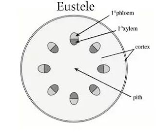
Eustele : if the stele is split into distinct collateral vascular bundle it is called eustele. It is a modification of the ectophloic siphonostele. E.g., Dicotyledons.

Atactostele: Differing from eustelic condition, the vascular strands are scattered. Such a stele has been named as atactostele by Esau. It occurs in monocotyledons.


Polycyclic stele: when the vascular tissue is present in the form of two or more concentric rings, such a stele is called a polycyclic stele. E.g., Pteridium aquilinium, Marattia. 


Sunday 17 March 2019

Equisetum


Equisetum
                                                                                                        Class Sphenopsida
                                                                                                        Order Equisetales
                                                                                                         Family Equisetaceae
                                                                                                          Genus Equisetum
Distribution
Equisetum is the only living representative of the family Equisetaceae. It is commonly known as Horse tail (aerial shoot with closely arranged whorls of branches look like a tail of a horse) or scoring rushes (due to sand papery  texture due to deposition  of silica).
The genus is represented by 32 species, found all over the world except in Australia and New Zealand.  Most species occur in the north Northern temperate zone but they are also known in Arctic, the southern and temperate and the tropics of both the hemispheres.
In India it is represented by about 7 species. E. arvense is a cosmopolitan species. E. debile  grows along the banks of sandy or swampy  soil and is found even in the Gangetic plains. Other Indian species are E. diffusum, E.ramossissimum, E. maximum, E. dubium.
Habitat
Equisetum grows under various habitats but thrives well under damp and amphibious conditions. Commonly they are found along the banks of rivers, streams ,( E. debile, E. protease) and swampy places (E.palustris). E. debile grows under hydrophytic as well as xerophytic conditions. E.avernse grows in open grasslands, railway embankments, exposed sandy and dry places. E. avernse is commonly called field horse tail.
According to Vogt,  few Equisetum species are used as ecological indicators, specially to indicate the mineral content of the soil in which they grow.  According to Benedict, certain Equisetum species accumulate minerals including gold and can therefore well be used to determine the presence of minerals in the soil.
Sporophyte
External morphology

The plant body of Equisetum represents the sporophyte generation. All species are well branched,  perennial herbs, containing a horizontal underground rhizome from which arise many roots towards lower side and many erect aerial shoots towards upper side.
Rhizome
Main stem is an extensively branched, sympodial, dark brown, rhizome. The rhizome penetrates deep into the soil. The rhizome presents a jointed appearance due to presence of distinct nodes and internodes.
On its nodes are present many scaly leaves. The scaly leaves are small, slender and united laterally with each other to form a sheet on the node.
The rhizome gives off  aerial as well as subterranean branches that alternate with the scale leaves.  A branch primordia, alternating with each leaf, is present on the node.
Aerial stem
From the rhizome, the aerial shoots arise towards upper side.  They are conspicuously jointed and are green, rough and stiff. They are abrasive due to deposits of silica.
The  jointed appearance is due to the presence of distinct nodes and internodes.
The nodes bear whorled leaves that are laterally fused at the base.  The internodes are long and hollow and are longitudinally rigid and grooved.
Each ridge corresponds to a leaf at the node above and the ridges in the successive internodes alternate with one another.
Another characteristic feature of the stem is the presence of an intercalary meristem that is present at the base of the internodes and just about the node. The meristematic region is protected by the leaf sheath. The activity of this meristem is responsible for the increase in length of the internode.
The aerial stem maybe branched or unbranched. In E. arvense the Ariel princess or profusely branched the branches arise in whorls at the node. They alternate with leaves.

In most spaces the aerial branches are distinguish as sterile and fertile branches.
Sterile branches are green with whorl of scaly leaves at each node. They are photosynthetic. At each node whorl of lateral branches arise, which are assimilatory in function. Each whorl bears an equal number of branches to that of leaves.
Fertile branches are unbranched, non chlorophyllous and terminate into strobilus at the apex.  In many species, the fertile branches  die as soon as the spores are released. In other species the fertile branch is green and after the cone sheds the spores, it behaves like the sterile branch

Leaves
Equisetum is micro fillers the leaves are simple irregular surface and extremely reduced in size they arise in viral at each node and their number varies with species.all the leaves are late relief use a sheet around the base of the internals the sheet and pointed teeth like tips. the leaves are mainly protective in function and in some species the leaves become dead and scale like at maturity the leaves do not perform any photosynthetic function at each node alternating with the leave there is a branch primordium the leave as a single median when they are usually without chlorophyll
Roots
The roots develop from the nodes of rhizome or from the base of the stem. They are long, slender, well branched and adventitious.
The roots arise indigenously from the pericycle. The roots are extensively branch and have a root cap at their tips

Tuesday 12 March 2019

General characters of Pteridophytes

Pteridophytes
Cryptogams can be classified into non vascular cryptogams and vascular cryptogams.  Since vascular system is absent in Algae, Fungi and bryophytes, they called as non-vascular cryptogams.

The term pteridophyta was first introduced by Haeckel in 1866. Pteridophytes
 (pteron-feather)possess vascular tissues and reproduce by spores they are called as vascular cryptogams. The reproduction by spores is the basis for placing pteridophytes under cryptograms.
Bryophytes and pteridophytes and gymnosperms are together called as archegoniates because of  presence of archegonium  in  all of this group of plants.

Sinnott in 1935 proposed a group of Tracheophyta to include the vascular plants i.e., pteridophytes and spermatophytes.

The pteridophytes appeared in early  Silurion period.  They formed a dominant Flora on the earth in the late devonian and carboniferous periods, about  35 million years ago.tree like massive arborescent forms flourished in the devonian and carboniferous periods but the present forms are small and herbaceous except the tree ferns. There are about 400 general and 10500 species of Living pteridophytes

Distribution and habitat
They grow in tropical and temperate regions of the globe. The plants grows luxuriantly in cool damp shady places. Some species flourish well in open,dry situations like Selaginella letridophylla, Equisetum arvers. A few pteridophytes are aquatic like Azolla and Salvinia. Some are epiphytic L.phegmaria, c.squarrosum, S.aregana.
All living pteridophytes are herbaceous except a few woody ferns like Cyathea.

The plant body:
The most characteristic feature of pteridophytes is the presence of independent sporophyte and gametophyte. The diploid sporophyte generation is dominant in pteridophyte in contrast to bryophyte. It attends a high degree of complexity with reference to external and internal organisation.
The sporophyte plant body is differentiated into root, stem and leaves for the first time in pteridophyte. Sporophyte shows such differentiation for the first time in plant Kingdom.
The primary root is short lived and it is replaced by permanent adventitious roots arising from the base of the stem from the nodes of aerial stem. The branching of roots is either monopodial or dichotomous. Roots shows diarch and exarch arrangement of xylem in the Stele.
The stem may be horizontal or erect. The creeping horizontal stem maybe subterranean or aerial or ephiphytic. The stem looks like a corm(Isoetes) or tuber(Equisetem).

The stem may be smooth or covered with hairs or scales. Branching maybe dichotomous as in Lycopodium or lateral as in  Polypodium.

The vascular system in stem varies in different species. Stele ranges from protostele to polycyclic condition. Xylem consists of tracheids only. Phloem is made up of sieve tubes but lack companion cells.  Vessels are reported in some members for example Selaginella and Equisetum.

Secondary growth is absent in majority of the living species, Isoetes being an exception.
In erect stem leaves are arranged around the stem in spirals or in pairs or in whorls. In rhizomatous stems the leaves arise from the upper side.

On basis of their size the leaves may be microphyllous or megaphyllous. In microphyllous leaves there is only midrib which is unbranched running from base to apex example Selaginella, Lycopodium. The megaphyllous leaves have a stout petiole and lamina with dichotomous venation. The leaves may be simple or compound. Young leaves of plants exhibit circinate vernation.
Internally the mesophyll may be un -differentiated as in microphyllous leaves (primitive character) or may be differentiated into palisade and spongy parenchyma as in megaphyllous leaves (advanced character).

Reproduction:
Vegetative reproduction takes place due to death and decay of the rhizome, tuber, Gemma and adventitious buds etc.
The plants reproduce asexually by means of spores. The sporophyte produces spores which may be  homosporous that is produce only one type of spores, (example Rhynia, Lycopodium, Equisetum) or heterosporous that is produces two types of spores the small microspore and larger megaspore, (example Selaginella, Marsilea).

The spores are produced in sac like structure called sporangia. Sporangia are usually born on leaf like structure called sporophylls on axils of the leaves. The sporophylls along with the sporangia are  scattered throughout the vegetative part of the plant or may be restricted to terminal parts.

In Equisetum and Selaginella the sporangia form distinct compact structures called strobilus or cone. In some forms the sporangia bearing special stalked structure called sporangiophore
 are arranged in whorls on the axis of the strobilus, e.g., Equisetum. The sporangia in some cases maybe produced within the bivalved specialised structures called the sporocarps example Marsilea, Salvinia, Azolla.
In true ferns (Filicales) the sporangia are usually born on group called sori. The sori are found on the margins or under abaxial surface of the leaves. The shape of sori maybe circular, linear or reniform. The sori are of three types based on the mode of development of sporangia in a sorus (Bower 1935).
1. The simple sorus:
All the sporangium in a sorus develop simultaneously and all of them mature together example Ophioglossum, Osmunda.

2.The gradate or basipetal sorus:
The sporangia develop in a basipetal manner that is the oldest sporangia is at the top
of the receptacle and the younger sporangium at the base example Dicksoria, Cyathea.

3.The mixed sorus:
The sporangium of different ages are intermixed in the same sorus and show no regular arrangement. The sporangia usually have long stalks and vertical annulus, example Pteris, Adiantum.

 The development of sporangium may be eusporangiate or leptosporangiate
Eusporangiate type :
The sporangium originates from a group of superficial initials known as sporangial initials this initials divide periclinally into outer primary cell walls and inner sporogenous cells
The primary wall cells develop into several layered wall of sporangium. The innermost wall layer functions of tapetum which forms a nourishing plasmodial fluid.
The wall of the mature sporangium maybe single layer due to degeneration of remaining layers, example Lycopodium, Selaginella, Equisetum.
 But in Psilotum and Tmesipteris the mature sporangial wall remains multilayered. The sporogenous cells divide meiotically and give rise to haploid spores. The spore output  is high that is 1000 to 25000 spores. Eusporangium is primitive.

Leptosporangiate type:
The leptosporangiate develops from a single superficial initial. The initial divides transversely into an outer and inner cell. The entire sporangium develops from the outer cells. The inner cell is responsible for the formation of stalk of the sporangium.
The mature sporangium is small and the sporogial wall  is single layer. The spore output is less that is 4-256 spores. The leptosporangiate is more advanced and found in Filicales (example Pteridium, Pteris, Marsilea).
Within the sporangium, the diploid spore mother cell undergo meiosis to form spores. The spores maybe homosporus (example Lycopodium, Equisetum) or heterosporous (example Selaginella, Marsilea). In  heterosporous type the smaller spores are termed as microspores and larger spores are termed as megaspores.
Spores are haploid and form after reduction division in the sporogenous cells of the sporangium.
Spore germination to multicellular gametophytic  body called prothallus.
The gametophytes are of two types:
Gametophytes that develop from homospores grow up on the soil surface and form independent plants. Such gametophytes are known as exosporic gametophytes example Lycopodium, Equisetum.
Gametophytes that develop from microspores and megaspores are for the most part retained within the spore wall. Such gametophytes are known as endosporic gametophytes example Selaginella,  Marsilea.  They live on food deposited in the spores.
Generally the prothallus are green, simple somewhat branched and aerial structures but in some genera such as lycopodium they are subterranean, well branched, tuberous, colourless.
The gametophyte or prothallus  bears the sex-organs, the antheridia and archegonia.
They are similar in structure to those of bryophyta
Generally the prothalli  of homosporous pteridophytes are monoecious and protandrous. Where as the prothallus of heterosporous pteridophytes are dioecious.
The antheridium
The antheridia may be embedded either wholly  or in part in the tissue of the gametophyte. The farmer are the embedded type example lycopodium selaginella acquisitive and the later are the projecting type example Pteris.
The antheridium is a spherical structure.  It has a sterile jacket that encloses a group of androcytes.  Each androcyte gives rise to a single motile antherozoid. The antheridia  are unicellular, uninucleate and biflagellate structures in Lcopodium,  Selaginella  but they are multiflagellate in Psilotum, Equisetum and ferns.
The archegonia
Each archegonium is a flask shaped structure consisting of a basal, swollen, embedded part,  the venter and a short projecting neck. The venter encloses the egg and venter  canal cell. Inside the neck are found 1-6 neck canal cells.
 Fertilization
 Fertilization takes place with the help of water and results in the formation of a diploid zygote antherozoids are attracted chemotactically  towards the archegonium.
Zygote
 zygote is the first cell of sporophyte and it is diploid.
Zygote develops into the embryo embryo maybe endoscopy where the effects of the embryo is directed inwards towards the gametophytic tissue ( Lycopodium,  Selaginella) or exoscopic in which the apex of the embryo is directed towards the neck of archegonium (Psilotum). It develops into a well developed sporophyte bearing roots, stem and leaves.
Pteridophytes show alternation of generations.

Wednesday 6 March 2019

Evolution of Sporophyte in Bryophytes


Evolution of Sporophyte in Bryophytes

The sporophyte of Bryophytes is a solid object, radially constructed and represents the diploid asexual generation in the life history of a plant. A product of diploid zygote, the sporophyte has its chief function, the production of spores which through a process of meiosis always possess the haploid chromosome number.
The sporophyte is incapable of self-nutrition and is wholly or partially dependent upon the parent gametophyte to which it is originally attached throughout its life. In form, it varies from only a spherical spore producing structure as in Riccia to an elaborate object differentiated into foot and capsule as in Corsinia to more elaborate structure consisting of foot, seta and capsule as in Polytrichum or Funaria
The foot functions as an anchoring and absorbing organ. The seta helps in conduction and aids in spore dispersal. Chlorenchyma tissue, stomata and air spaces for efficient food synthesis. Elaters, Operculum, Peristome for dehiscence of capsule and spore dispersal. Columella for storage of water and soluble food (in mosses) and for mechanical strength (as in Anthoceros)

Two theories have been put forth to explain the process of evolution of the sporophyte.

Theory of Progressive Sterilization              

This theory is was proposed by Bower and supported by Cavers, Campbell and Smith. According to this theory, the most primitive sporophytes in Hepaticae occurs in the genus Riccia. Such sporophyte, according to some bryologists, are the nearest hypothetical ancestors or represents the actual ancestors of more highly evolved group of plants.
The sporophyte of Bryophytes, according to their complexity of structure may be arranged in a series between the simplest and the most elaborate. This series starts with simple sporophyte of Riccia, runs through that of Marchantia, Pellia, Anthoceros and finally culminates in the highly complex sporophyte of Funaria and Polytrichum.
According to Bower, this series runs in the upward direction. And illustrates a natural advance in the progressive elaboration and complexity of the sporophyte. It is based on the fundamental principle of ‘Progressive sterilisation of the potentially fertile cells (sporogenous tissue)’. Instead of forming spores, the sterile cells develop into elaters, nurse cells and elatophore in different genera. These sterile cells are put to other uses such as nutrition, support, dehiscence, dispersal, etc.
The progressive sterilisation from Riccia to Polytrichum occurred through following stages:

Step – I : The simplest known sporophyte among bryophytes is that of Riccia. It consists of a spherical capsule and seta and foot are absent. The zygote divides by a transverse and then by a vertical division to form a 4- celled embryo. This becomes 20-30 celled by further division.
Periclinal division at this stage differentiates the single layered amphithecium from an inner multicellular endothecium. The amphithecium forms the   layered capsule wall. The endothecium transforms into archesporium, which divides and redivides to form the  fertile sporogenous tissue.
The entire sporogenous tissue is fertile and is forms the spores. There is large output of sores and no or very little sterilisation of the fertile cells. The entire embryo forms the spore producing capsule.
In Riccia crystalline some of the sporogenous cells fail to form spores, but instead they form sterile nutritive cells.

Step – II: In this step, further sterilisation of the embryonic tissue led to the formation of a basal sterile foot of few cells. The Oospore divides into a hypobasal and an epibasal cell. The hypobasal cell gives rise to the foot. The epibasal cell form an outer amphithecium and inner endothecium. The amphithecium gives rise to a single layered jacket and the endothecium differentiates into fertile sporogenous tissue and sterile nurse cells. The nurse cells are long, elater-like, but lack characteristic thickening.
Thus, in Corsinia, the sterilisation has gone a step further and resulted in nurse-cells and foot.


Step – III: Further sterilisation is seen in Sphaerocarpos, where the sporophyte has a sterile bulbous foot and a narrow seta, in addition to fertile capsule. The amphithecium forms the single layered jacket of the capsule and the endothecium forms the sporogenous tissue and the sterile nurse cells.


Step – IV: Targionia exhibits a further step in progressive sterilisation of the potentially sporogenous tissue. The sterile region in this genus consists of a broad foot, a well-developed seta and a large number of elaters with 2 or 3 spiral thickenings.
The amphithecium give rise to a single layered jacket of the capsule. About half of the endothecial cells gives rise to fertile sporogenous tissue and the remaining half form the sterile elaters.


Step – V: This step is exemplified by the sporophyte of Marchantia. The lower half or hybobasal cells of the embryo forms the foot and seta. While, the upper half or epibasal cells forms the capsule. The amphithecium gives rise to a single-layered capsule wall or jacket.
The endothecium forms the sporogenous tissue. Half of the  sporogenous tissue forms spores and the remaining half of the tissue elongate, develop spirally thickened bands on their walls and becomes elaters. The elaters are hygroscopic and help in the dispersal of spores.
Thus, the capsule of Marchantia has specialised both as a spore  producing and spore dispersal body. It illustrates a step further in the progressive sterilisation of the sporogenous tissue and consequent elaboration of its sporophyte.

Draw Marchantia Sporophyte from the Record

Step – VI: Further sterilisation of the sporophytic tissue can be seen in Jungermaniales (e.g., Pellia, Riccardia). The hypobasal half of the zygote takes no part in the development of the sporophyte. The entire sporophyte including the foot and seta is developed from the epibasal half.
Epibasal half differentiates into an outer amphithecium surrounding the inner endothecium. The amphithecium forms the  2or 3 layered capsule wall. The endothecium in whole develop into sporogenous tissue.
The sporogenous cells at the base of the capsule remain sterile. These sterile cells elongate considerably and develop spiral thickenings on their walls to become elater-like. This elater-like cells attached at their lower ends to the cavity of the ca psule is called the basal elaterophore.
In addition, some other dispersed cells in the rest of the sporogenous tissue form elaters which remain unattached.
Only small percent (10%) of sporogenous tissue forms the spores.

Step – VII : this step is illustrated by Anthoceros sporophyte. There is complete sterilisation at the centre. The entire endothecium remains sterile and from a central column called the columella, which is useful for storage of water and food and for mechanical support.
The amphithecium forms the capsule wall and sporogenous tissue. Thus sporogenous tissue arises from the innermost layer of the amphithecium. The wall of the capsule is multilayered consisting of epidermis, stomata , the photosynthetic chlorenchyma .
Even the sporogenous tissue differentiated into fertile spore mother cells and sterile pseudo elater mother cells. The pseudoelater mother cells forms the pseudoelaters as they do not possess spiral thickenings.
Thus, the spsore producing area is greatly reduced and the sporophyte shows a great degree of nutritional independence due to photosynthetic tissue.

Draw Anthoceros sporophyte from the Record

Step – VIII : The highest degree of sterilisatio of the sporogenous tissue is found in the class Bryopsida (Mosses) e.g., Funaria and Polytrichum. Major portion of the embryo remains sterile to form seta and foot. The amphithecium becomes differentiated into the epidermis, the photosynthetic tissue and air space or lacunae.
Except the superficial layer there is complete sterilisation of the endothecium to form the central columella which is continuous right up to the top of the capsule. The archesporium arises from the outermost layer of the endothecium. It is thus extremely reduced and consists of a single layer of cells and confined to the theca region only.
The sterilisation of archesporium towards the base results in the increase in size of the photosynthetic tissue in the apophysis region. The fertility is arrested towards the top due to specialisation of this region for spore distribution because of the formation of peristome, operculum and annulus.
Thus, moss capsule is mechanically more efficient in dispersal of spores, consequently the archesporium is much more reduced.
Bower’s theory of sterilisation offers a plausible explanation of the evolution of the sporophyte in the upward direction.


Theory of Regressive Evolution or Reduction Theory
This is also known as theory of progressive simplification or reduction theory
This theory was proposed Kashyap and supported by Church, Goebel and Evans.

According to this theory Riccia sporophyte is the most evolved or the advance one formed due  to reduction as a result of progressive simplification . They think that the primitive sporophytes is  like that of Mosses (e.g. Funeria and Polytrichum)from which the sporophyte of  Marchantia, Jungermanniales and Anthocerotales have been evolved by  reduction of the tissues.
Church suggested that the hypothetical ancestors of bryophytes had foliose sporophytes with complex assimilatory tissue and  epidermis with functional stomata.  From such a plant,  evolution progressed  regressively as follows:
1. The erect leafy sporophyte became permanently attached to the gametophyte and gradually lost its leaves.
2. Reduction of the green photosynthetic tissue in the capsule wall.
3. Associated with the above is the disappearance of stomata and intercellular spaces.
4. The multilayered sterile jacket of the capsule ( Funeria, Anthoceros)became single layered by (Riccia, Marchantia)by reduction
5. Gradual elimination of the anchoring tissues like foot, seta, apophysis and the conducting tissues like columella
6. Archesporium formation shifted from amphithecium to endothecium.
7. Progressive increase in the fertility of the sporogenous cells these changes eliminated the presence of sterile cells and relatives in the capsule.

Monday 4 March 2019

Polytrichum

Bryopsida
Distribution and Habitat:
The moosses are the higher Bryophytes. They are world wide in their distribution.
Occur in almost all situation were life is possible. The only exception is the sea. The
leafy plant body of a moss plant is better adapted for a life on land than the thallus of
liverworts. However, they flourish the most wet and humid region, such as, moist
mountain forest of tropics and sub tropics. A few are aquatic. A few , such as,
Sphagnum grow in bogs. Theya re found in tempetate and arctic tundras. They grow
on soil,fence, rocks tree trunks.


Polytrichum
Division: Bryophyta
Class:      Bryopsida
Sub:Class: Bryidde or Eubrya
Order :    Polytrichales
Family :   Polytrichales
Genus  : Polytrichum
Distribution and Habit:
The genus Polytrichum is represented by about 100 species which are widely distributed all
over the world. They are mainly distributed in cool temperate and tropical regions of the world.
Polytrichum mostly prefer moist and shady places. The plants grow on wet sandy banks of
rivers and ponds, on branches of trees under shade (Epiphytes) and on rocks and cliffs,
on bogs. They often form a green carpet on moist and shaded walls.
In India, the genus is represented by five species found in Himalayan regions –
P. densifolium, P. juniperinum, P. xanthopilum, P. alpinum, and P. formosum.


Gametophyte
External Morphology
Polytrichum gametophyte has two stages 1. Protonema stage 2. Adult gametophyte stage.


Protonema
Spores on germination produce creeping, green, branched and filamentous protonema.
Protonema represents the temporary junvenile phase of the gametophytic generation.
The growth of protonema is apical. Two types of filaments grow on the protonema,
upward and green chlorenematous filaments and colourless rhizonematous filaments.
The rhizonema filaments are meant for attachment. On upright chloronema filaments
buds develops. Each bud develops into an erect leafy gametophore.
Gametophore
The gametophore is a erect leafy shoot. The gametophore is differentiated into two parts :
a horizontal underground rhizome and an erect leafy shoot.


Rhizome
It is underground, horizontal growing portion of the gametophore. The rhizome bears leaves
and fluffy rhizoids.
The leaves are small, scale-like, usually brown or colourless. They are arranged in three
vertical rows (1/3 phyllotaxy).
The rhizoids arise from the base of the rhizome. Rhizoids are long, thread like, multicellular,
branched with oblique septa. Rhizoids are inter woven to form a wick like structure.
Rhizoids absorb water not only through their surface but also hold water externally
by capillary action as in the wick of a lamp. Because of this character Polytrichum can
withstand relatively dry habitats.
Buds and gemmae present on the rhizoids help in vegetative propogation.


Ariel Leafy Shoot
The leafy shoot is an erect axis arising from the horizontal rhizome. It is the most conspicuous
part of the plant. It grows to a height of about 15-20cm.  In P. commune, the leafy shoot grows
upto 45 cm in height
Each leafy shoot consists of a central axis and many lateral expansions called leaves. Typically,
the erect leafy axis is unbranched. The central axis bears two kinds of leaves, the scale leaves
and foliage leaves. The scales leaves are produced on the lower portion of the central axis. The
foliage leaves are large and are arranged spirally on the central axis with a divergence of 3/8.
Each leaf consists of two parts, a broad colourless, membranous sheathing leaf base and a narrow,
brown or green lanceolate limb.
The limb consists of a broad dark green midrib and rudimentary wings. The wings on both sides
of the midrib are thin. The leaf margin may be entire or toothed.
The branches arise from the primordial present below the leaves. The shoot grows by means of an
apical cell with three cutting faces.


Internal structure
Rhizome
The rhizome is roughly triangular in outline with rounded corners. The transverse section shows
three regions an eipidermis, cortex and central cylinder.


Epidermis
It is outermost layer of the rhizome. It is single layered. The epidermal cells are thick-walled due
to deposition of suberin. Several epidermal cells give rise to rhizoids. Stomata are absent.


Cortex
The epidermis is followed by cortex consisting of 3-4 layers of thin walled cells. The cortex is
interrupted by three hypodermal strands, which extend radially from the periphery towards the
centre. They are composed of living prosenchymatous cells, which contain starch grains.
The hypodermal strands gradually narrow down towards the centre of the rhizome, and are
connected inward with thin walled cells. These thin walled cells together with hypodermal
strands are called radial strands.

Inner to the cortex, there is a layer of radially arranged large cells. The radial and horizontal
walls of these cells have suberized thickening. This layer can be compared with the endodermis
of higher plants. This layer is not continuous but separated by radial strands.
Central Cylinder:
It is the central, compact, 3-lobe mass of tissue forming the core of the rhizome.
Pericycle: In some species, a 2-3 layers of thin walled pericycle is present between the
endoermis and the central cylinder. It is not continuous and is absent in the region of furrows
where the centre of the bay is occupied by the lepoids.
Leptoids:
In the furrows of the interrupted pericycle, is present a group of polygonal proteinaceous
cells. These cells are large, elongated sieve tube-like and termed as leptoids. Collectively the
three groups of leptoids, which more or less resemble the sieve cells of the vascular plants,
constitute the leptom.
These are living cells with oblique end walls and are connected with each other through
plasmodesmata. The leptom is the food conducting system.
Amylom:
The inner most layer of leptoids is seperated from the central cylinder by a single layer
of parenchymatous cells containing starch. This layer is called amylom. The amylom
seperates the leptom from the central trilobed hydrom.

The central cylinder consists of two kinds of elements, the stereids (or sclereids) and hydroids.
The central mass mainly consists of the stereids. These arre thick-walled, elongated cells with
oblique end walls. Collectively the stereids constitute the stereom. It functions as the supporting
tissue.
Interspersed among the stereids are the empty elements in groups of 2 or 3. These are termed as
hydroids. Collectively the hydroids constitute the hydrom. They function as the water conducting
tissue and is equalent to the xylem of the vascular plants.
The stereom and the hydrom together constitute the hydrom cylinder.


T.S of Aerial Stem:
The cross section of the erect stem has an irregular outline due to attachment of leaves.
It is differentiated into epidermis, cortex and central cylinder.
Epidermis:
It is single layered, usually not well defined.
Cortex:
It is differentiated into outer thick walled cortex and inner thin walled cortex. the leaf traces
pass through the cortex and join the central cylinder interrupting the pericycle.
The inner cortex is followed by endodermis. Inner to endodermis is a zone composed of
thin-wlled, sieve tube like cells. It is known as leptom mantle. It performs the function
of phloem.
Inner to leptom mantle is a narrow zone consisting of a single layer, sometimes of two
layers, of cells containing starch. These cells have dark-brown suberized walls. This
zone is called the hydrom sheath or amylom sheath.
Central Cylinder
In the centre of the stem is a compact mass of thick walled cells constituting the
hydrom cylinder. It consists of two kinds of cells, the stereids and hydroids.
Stereids are thick-walled supporting cells constituting the major part of the hydrom
cylinder. Interspersed among the stereids are the thin-walled elongated empty cells
in groups of 2 or 3. These are the hydroids concerned with water conduction.
Surrounding the hydrom cylinder is a zone consisting of two or three layers
of thin-walled cells devoid of contents (empty)

Anatomy of Leaf
The cross section of the leaf shows a broad midrib flanked by a narrow wing or lamina.
The midrib makes up most of the width of the leaf. it is multistratose (several cells thick)
in the centre. Gradually, it thins towards the margins and finally merges into the wing
 or lamina on either sides.
The lower epidermis is single layer consisting of regularly arranged large cells with
their outer walls thickened.
Within the lower epidermis, the midrib shows one or two layers of sclereids cells with
extremely thickened walls and narrow lumen.
There is narrow and interrupted band of similar cells on the upper (adaxial) side.
Between these two bands of sclereids, the midrib consists of thin-walled parenchyma cells.
The midrib lacks the upper epidermis. It is replaced by a layer of large, thin-walled cells.
These cells bear the rows of green or dark green cells. These cells are 4-7 cells high,
standing vertically parallel to each other. These cells are called the lamallae and contains
chloroplast.
The lamallae besides being photosynthetic also hold water due to capillary force. The
terminal cells of the lamallae are colourless and enlarged. In P. commune it is bifid and
in P.junniperinum it is papillose.
The adjoining terminal cells almost touch each other, thus providing a functional equivalent
 of the upper epidermis. It is therefore, termed as pseudo-epidermis.

Reproduction:
Vegetative Reproduction:
The gametophore of Polytrichum is rhizomatous. Death or decay of the intervening rhizome results
in the establishment of seperate free living plants.
Sometimes, vegetative buds or bulbils developed on the rhiziods also helps in vegetative propagation.

Sexual Reproduction:
All species of Polytrichum are dioecious. The antheridia and archegonia are borne on different
gametophores. The sex organs are borne in groups at the tip of the main axis of the gametophore
which is unbranched.

Antheridial Head:.
Antheridia are borne in groups at the apex of the male plant. The leaves surrounding the antheridia are called the perigonial leaves. They differ from the vegetative leaves in form and colour. The perigonial leaf is comparatively shorter, red-brown or dull-red in colour. The leaves consists of a
broad expanded leaf base terminating in a short bristle point.
The perigonial leaves lie close to each other forming a rosette around the antherida  superficially resembling a miniature flower (moss flower). The apical of the male gametophore is not used in the
formation of antheridia. So, the axis may grow out through the antheridial head and produce antherida year after year. Such growth pattern is called as proliferation.
Intermingled with the antheridia in the antheridial head are multicellular hair-like structures called
the paraphyses. They are simple, filamentous structures consisting of a single row of uniform cells.
Some may have broad spathulate tips.
Antheridium
The mature antheridium consists of an elongated, club-shaped body and a short multicellular stalk.
The body of antheridium has a single layered jacket surrounding a central mass of primary androgonial cells.
Each primary androgonial cell divides repeatedly to form several androgonial cells. The last generation of the androgonial cells are called androcyte mother cells. Finally the androcyte mother cells divide producing androcyte. Each andorcyte metamorphoses into antherozoid.
An operculum is present at the tip of the antheridium, consisting of  4-5 distal tiers of small cells with markedly thickened walls.
The antheridium ruptures at the distal end due to absorption of water.The biflagellate antherozoids comes out. Each antherozoid has a coild body witha cytoplasmic vesicle attached at its posteriod end.

Archegonial Head:
The archegonia occur in a cluster at the tip of female gametophore. The leaves surrounding the archegonia are called the pericheatial leaves. The pericheatial leaves overlap at the top of the archegonial cluster to form a bud-like structure called the pericheatum.
Intermingled with the anrchegonia are the paraphyses. Each paryaphysis is a filamentous structure consisting of a row of uniform cells.
The apical cell of the female gametophore itself functions as the archegonial initial. Consequently further growth of the female gametophore is not there.
Structure of Archegonia
The mature archegonium is flask shaped structure consisting of a very long neck, venter and massive stalk.
The wall of venter is two cell thick. The venter cavity contains a single spherical egg and a ventral canal cell just above it. The long, narrow neck consists of six vertical rows of neck cells enclosing neck canal cells (about 13 or more).
The venter canal cell and neck canal cell in the mature archegonium degenerate forming a mucilaginous mass .It absorbs water and swells forming a passage way leading to the egg in the venter.

Fertilisation:
The perigonial bracts surrounding the terminal antheridial cluster lie close together and overlap forming a shallow cup-like structure. It serves as a splash cup. Falling rain drops from a height would result in splashes coated with sperms travelling considerable distance.
Splashing drops containing brings sperms to the archegonial cluster. The sperms are released into archegonia. A single antherozoid fuses with egg to accomplish fertilisation. The fertilised egg secretes a wall around it to become an oospore.

Sporophyte
The diploid zygote is the first cell of the sporophyte.
Located in the venter cavity, it increases in size and divides by a transverse wall into an upper epibasal cell and a lower hypobasal cell. Each cell divides further and forms an apical cell with two cutting faces.
The apical cell formed in the hypobasal cell gives rise to foot, while the apical cell of the epibasal cell gives rise to seta and capsule.
The cells destined to form capsule divide periclinally and form an outer amphithecium and an inner endothecium. The amphithecium gives rise to the jacket of the capsule, whereas the endothecium gives rise to columella and the archesporium.
Along with the development of sporophyte, the wall venter divides forming 4-6 cells covering called the calyptra. It completely encloses the developing embryo. The calyptra is ruptured by the continuous growth of the sporophyte. The upper part of the calyptra is carried upward on the top of the capsule as dry hairy hood. Due to this hairy calyptra covering the mature capsule Polytrichum is commonly known as Hairy Cap Moss.

Structure of the Mature Sporophyte
The mature sporophyte or sporogonium of Polytrichum is differentiated into foot, seta and capsule.
Foot:
It is dagger-shaped structure buried deep in the tip of the female gametophore. It is composed of thin-walled parenchymatous cells. It functions as an anchoring and absorbing organ.
Seta:
The seta is a long, slender, stalk-like structure present between the foot and the capsule. Structurally it consists of an outer layer of thick walled cells constituting the epidermis. Epidermis is followed by a sclerenchymatous hypodermis. Next to hypodermis is the cortex consisting of green, thin-walled parenchymatous cells with intercellular spaces. Forming the core of the seta is a well developed central strand.
The main function of the seta is support and conduction of the water and mineral salts, raise the capsule to the required height.

Apophysis:
At the base of capsule, the seta is considerably swollen to form a bulbous, sterile structure called thee apophysis. It is demarcated from the Capsule  by a disntict groove. Structurally it consists of a single layer of epidermis continuous with the epidermis of seta below. It has stomata. Next to epidermis is the chlorenchyma consisting of a mass of loosely arranged thin-walled parenchymatous cells containing chloroplasts.
The hypodermis is absent in the apophysis region. In the centre is the conducting strand continuous with the columella above.

Capsule:
The capsule is long, erect and angular in shape. It is differentiated into two regions, (i) the lower, large spore bearing portion forming the body of the capsule called the theca and (ii) a sterile, conical portion forming the lid or operculum.

Theca:
The fertile part of the capsule extending from the apophysis to operculum is called as theca.
The theca consists of several cell layers called the capsule wall. The outermost layer of the wall is epidermis with compactly arranged cells. The epidermis of the capsule region lacks stomata.
Next to epidermis is the chlorophyll tissue typically 2-celled in thickness. It consists of thin-walled parenchymatous cells containing chloroplasts.
Inner to the chlorenchymatous tissue is the outer air space or lacunae. It is traversed by short filaments of green cells called the trabeculae. The trabeculae connect the theca wall layer with the
outer wall of the spore sac.
The outer lacunae is followed by spore sac. The spore sac contains the fertile archesporial tissue. The archesporium originates as a single layer from the outer layer of the endothecium. Later it divides to form 4-6 layers of sporogenous tissue. All cells of the sporogenous tissue are fertile. They divide and form spore mother cells. The spore mother cells undergoes meiosis to form 4 haploid spores or meiospores.
Internal to the spore sac is inner air space or lacunae. It is also tranversed by trabeculae which connect the two cell layer thick inner wall of the spore sac on the outer side  to the central columella on the inner side. Thus, the spore sac is bounded by air space or lacunae  both at its outer and inner face.
The central part of the capsule is occupied by a solid core of sterile tissue forming the Columella. It is made up of parenchymatous cells and is continuous with the central strand of seta below and extends upto the epiphragm of the operculum above.

Operculum or Lid:
It is the terminal, conical portion of the capsule, which forms a cap or lid like structure at the apex of the theca.
The operculum is delimited from the theca region by constriction made of 2-3 layers of  elongated cells forming the rim or diaphragm. The annulus is absent.
Closing the mouth of the theca is a thin, shield-shaped pale membranous structure called the epiphragm. It stretches like the tymphanum of a drum across the mouth of the theca.
Arising from the periphery of the diaphragm is a ring of 64 short, stout pyramidal, teeth like solid structures called the peristome. Each peristome tooth is composed of curved crescent-shaped, fibre-like cells. At their tips, the teeth are joined to the margin of the epiphragm. They do not exhibit hygroscopic movements.

Dispersal of Spores
In the mature capsule, the central, non-sporogenous tissue degenerates. The spores come to lie in the hollow capsule. The calyptra falls off. The exposed mature capsule begins to dry. Minute holes appear between the successive peristome teeth in the margin of the epiphragm by the drying up of cellls between them.
The minute spores are dispersed, by censer mechanism as the capsule sways in the wind.
Spore:
The small yellow spores have a smooth surface. The spore wall is differentiated into outer exospore and the inner endospore. The reserve food is in the form of globules in the spore cytoplasm.

.