Thursday, 25 October 2018

Polysiphonia


Polysiphonia
                                                                                                            Class: Rhodophyceae
                                                                                                            Sub-class: Florideae
                                                                                                            Order:       Ceramiales
                                                                                                            Family :Rhodomelaceae
                                                                                                            Genus  :Polysiphonia

Distribution and Habitat:
            It is a common red algae with about 200 species on sea coasts. It is abundant in Atlantic and Pacific oceans. Most of them are found in littoral zone in tidal marshes, brackish estuaries and tide pools frequently growing as epiphytes on rock weeds.
            The genus is represented in India by about 16 species, which occur in the southern and western coasts. P. platycarpa, P. ferulaceae, P. urceolata, P. variegata are common India species.
            P. variegate inhabits polluted water near estuaries and frequently found on roots of mangroves. P. urceolata is an epiphyte on Laminaria. P. fastigata is found on the fronds of a brown sea weed Ascophyllumnodurum and occasionally on Fucus.

Thallus Structure:
            Polysiphonia has a filamentous thallus which is generally brownish red to purplish red in colour. The filaments branch and re branch several times giving the plant body a beautiful, delicate, feathery appearance.The thallus is attached to the substratum in water by a holdfast.
The plant body is heterotrichous consisting of an erect or projecting system and a filamentous prostrate system.
Basal Prostrate System:
            This creeps over the substratum. It is anchored to the substratum by means of thick-walled, elongated, unicellular rhizoids arising from the  peripheral cells facing the substratum. The distal ends of rhizoids expand to form flat irregularly lobed attachment pads or discs. The creeping filaments function as a means of perennation.


Upright or Vertical Filaments:
            These arise from the creeping filaments. They may attain a height of 25-30 cm. These filaments remain free-floating in water. The branching is lateral and branches are of two kinds, long and short.
            The short branches are of limited growth and are known as trichoblasts. The trichoblasts are colourless, hair-like and forked. They are borne on the long, erect branches of unliminted growth. The trichoblasts usually bear male and female reproductive structures. Trichoblasts are usually deciduous and are shed annually in the perennial species before winter.
            The long branches arise from the basal cells of short branches. They show unlimited growth due to the activity of apical cell and do not bear reproductive structures.
            The main filament and long branches, each consists of a system of parallel filaments. These are called the siphons. There is one central filament termed the axial or central siphon. The central siphon is surrounded by peripheral filaments called the pericentral siphons. The number of pericentral siphons varies from 4-20 (P. elongate – 4, P. spiralis – 5, P. variegate – 6).
            Such a thallus having central siphon surrounded by many pericentral siphons is called as Polysiphonous. This algal genus thus derives its name from the polysiphonous nature of its thallus. The cells of the central and pericentral siphons are inter connected through pit connections, a feature characteristic of the red algae.
            The long branches are polysiphonous or multiaxial. The short branches or trichoblasts are made up of only central siphons, and the pericentral siphons are absent, so they are monosiphonous.
            Because of nearly the same length of the cells in the central and pericentral siphons and the regular manner in which the cells are arranged, the main axis appears to be differentiated into nodes and internodes.
Cell Structure:
            The central and pericentral siphons are made up of elongated, cylindrical cells consisting of the cell wall enclosing the protoplast. The cell wall is thick and is differentiated into two layers. The outer layer is made up of pectic materials and the inner of cellulose.

            The centre of the cell is occupied by a vacuole bounded by tonoplast. The cytoplasm is restricted to the periphery of the cell. The peripheral cytoplasm encloses a single nucleus and a number of discoid chromatophores. Chromatophores are devoid of pyrenoids.


The photosynthetic pigments located in chromatophores are chlorophyll – a, Chlorophyll – d, α,β carotene, biliproteins – r phycoerythrin and r- phycocyanin and a few xanthophylls.
The reserve food materials are floridean starch and floridoside.
Growth:
            The thallus grows by means of a dome shaped apical cell. It is situated at the  extreme tip of the naked part of the central siphon. The apical cell by transverse division forms a series of segments parallel to its posterior face. These segments elongate to form the axial siphon.
            Some of the sub-terminal axial cells divide periclinically and form a definite number of pericentral cell around the axial row of cells. Both central and pericentral cells elongate into their respective siphon with pit connections.
Reproduction:
            In life cycle of Polysiphonia, three types of plants are found. They are :Gametopyte, the Carposporophyte and the tetrasporophyte.

Gametophyte:
            It is a free living haploid plant. It is concerned with sexual reproduction and bears sex organs.
Carposporopohyte:
            It is diploid plant and develops from the zygote. It remains attached to the female gametophyte plant, on which it is parasitic. It reproduces asexually by producing diploid spores called the carpospores.
Tetrasporophyte:
            The diploid carpospores germinate to give rise to the tetrasporophyte plant. It is an independent diploid plant. It reproduces asexually by producing the haploid tetraspores.
            The haploid gametophytes and diploid sporophyte plants are similar in their morphological structure, but differ in producing the reproductive organs.


Gametophyte:
            The free-living Polysiphonia plant is a haploid gametophyte. It undergoes sexual reproduction which is an advanced oogamous type. The gametophyte plant is heterothallic or dioecious i.e., male sex organs and female sex organs are borne on different plants called the male gametophyte and female gametophyte resepectively. The male and female plants are morphologically similar.
Male gametophyte:
The Male sex organs, spermatangia or antheridia develop on fertile trichoblasts present on tip of male gametophytic plant. The male trichoblast  when only 2-3 celled, divides dichotomously. In most of the species one branch remain sterile and the other bears spermatangia. In some species both branches become fertile.
The cells of fertile uniaxial trichoblast  divide periclinally to form  pericentral cells. The  pericentral cells form the  spermatangial mother cells on outer side. Each spermatangial mother cell cuts off 2-4 sporangia on outer side. The complete structure makes cone shaped cluster of spermatangia.
The mature spermatangium is a globular or oblong, unicellular structure. Its cell wall is differentiated into three layers – inner refractive , middle gelatinous and outer thick layer. The uninucleate protoplast of spermatangium forms a male gamete or spermatium. The spermatium is non-motile and is released through an apical pore.


Female gametophyte:
The female sex organ is called carpogonium. The carpogonium develops on trichoblast on female gametophyte plant.  The trichoblast initial arises from a cell, 2-4 cells behind the apical cell. It develops into 5-7 celled female trichoblast.
The 3 lower most cells of the female trichoblast divide vertically to form an ensheathing layer of pericentral cells. One of the pericentral cell on the adaxial surface (facing the axis side) functions as supporting cell.
The supporting cell cuts of a small initial cell at its free end known as procarp initial. The initial divides to form a small, curved four-celled branch called the carpogonial filament or procarp.
The terminal cell of the carpogonial filament functions as carpogonial mother cell. The carpogonial mother cell gets modified into the carpogonium. The carpogonium has a basal swollen portion having female nuclei or egg and an upper elongated, slender portion called the trichogyne. The trichogyne functions as a receptive organ

Meanwhile, the supporting cell cuts off two sterile cells, one towards its base called the basal sterile filament initial and one on the lateral side called the lateral sterile filament initial.
At this stage the carpogonium is ready for fertilisation.


Fertilisation:
The liberated spermatia are carried passively by the currents of sea water. As they reach the carpogonium, one of them adheres to trichogyne. At the poing of contact the common walls between them dissolves. The male nucleus then enters the trichogyne and passes down and reach the eagg. The male nucleus and the egg nucleus fuse and a diploid zygote is formed.

Post- Fertilisaion Changes:
After fertilisation many changes takes place within and around carpogonium. The two-celled lateral sterile filament divides and becomes 4-10 celled. Then basal sterile filament initial divides to form a 2-celled basal sterile filament. The sterile filaments are nutritive in function.
Meanwhile, the supporting cell divides transversely to form an auxillary cell at its upper end. It lies between the supporting cell and the carpogoniium. It has a haploid nucleus. Soon, the auxillary cell establishes a tubular connection with the carpogonium.
The diploid zygotic nucleus of the carpogonium dividies mitotically into two daughter nuclei. One of the daughter nuclei migrates into the auxillary cell through the tubular conncetion. Other daughter nuclei remain within the carpogonium. The haploid nuclei of the auxillary cell degenerate at this stage.
After, the migration of the diploid nucleus into the auxillary cell, the carpogonial branch gradually begins to degenerate.
The migrated nucleus present in the auxillary cell divides mitotically into two daughter nuclei. One of these remains in the auxillary cell and the other migrates into a small lateral outgrowth arising from the upper side of the auxillary cell. This small outgrowth on the auxillary cell is called as gonimoblast initial.
The gonimoblast initial cuts off a number of cells. Every time a new cell is cut, the zygote nucleus divides and its derivative enters the newly formed cells. Thus, gonimoblast initial grows into a number of short threads, the gonimoblast filament.
The terminal cell of the gonimoblast filament becomes swollen and it develops into a pear-shaped carposporangium. The diploid protoplast of the carposporangium develops into a single, diploid carpospores.

With these development taking place, the supporting cell, the auxillary cell and cells of sterile filaments fuse resulting in a large, irregularly shaped structure called the placental cell. The placental cell provides nourishments to the growing carposporophyte.
Meanwhile, the pericentral cells of the female trichoblast adjacent to the supporting cell grow into outgrowths known as the enveloping threads.
The enveloping threads finally develop into an urn-shaped envelope or sheath around the developing frutification. This sheath is called the pericarp. It consists of two layers and has a wide aperature, the ostiole at its distal end.
The entire structure consisting of the placental cell, gonimoblast filaments bearing carposporangia and the surround pericarp is called the cystocarp. It is partly haploid and partly a diploid structure.


Carposporophyte or Cystocarp:
The post-fertilisation fructification respresenting the diploid phase and second individual in the life-cycle of Polysiphonia is called as carposporophyte. It remains attached to the female thallus of Polysiphonia and thus lives parasitic on it.
It is urn-shaped and protected by a two layered wall called the pericarp. The pericarp is a haploid structure. The cystocarp has an opening on its top called the Ostiole. The cystocarp contains a placental cell, gonimoblast filaments and carposporangia.
Each carposporangia produces a single, uninucleate, diploid carpospores. Carpospores are liberated through the ostiole and they are carried by the water currents.

Germination of Carpospore:
On coming in contact with substratum diploid carpospores secrets a wall around it and attaches itself to the substratum. It divides transversly and forms a smaller lower cell and a larger upper cell. Each of these again divide transversely forming a four-celled small filament.
The basal cell of the filament is called the rhizoidal cell. It is colourless, elongated and form the rhizoids. The end cell of the filament is dome shaped and functions as the apical cell. The lower axial cell divide vertically to cut off the pericentral cells. In this way a full-fledged tetrasporophyte is formed.
Tetrasporophyte:
It is an independent, diploid plant developed from the carpospores. It is morphologically similar to the haploid gametophyte. It consists of a central siphon encircled by the perincentral silphons. The thallus is laterally branched.

Asexual Reproduction:
The tetrasporophyte plant reproduces asexually by means of non-motile haploid spores called tetraspores. Tetraspores are produced within spherical, sac-like diploid structures called the tetrasporangia.
The tetrasporangia are developed from the pericentral cells. Only one of the pericentral cell in each transverse tier produces a tetrasporangium. The fertile branches bearing the tetrasporangia become swollen and twisted.
The fertile pericentral cell is smaller in size than the other cells in the same tier. This cell divides by a vertical wall into two cells, the outer and inner.
The outer cell again divide to form two cover cells. The inner cell functions as the sporangial mother cell. The sporangial mother cell divides by a transverse wall into a lower stalk cell and an upper tetrasporangium cell.
The tetrasporangium cell increases considerably in size. It has a diploid nucleus. The nucleus undergoes meiosis forming four haploid daughter nuclei.
This is followed by cleavage of the cytoplasm from the periphery towards the centre resulting in formation of four uninucleate meiospores. These four meiospores are arranged tetrahedrally in the tetrasporangium, hence called as tetraspores.
When tetraspores reach maturity, the sporangial wall ruptures and two cover cells move apart longitudinally. Thus, the tetraspores are liberated.
The liberated tetraspores germinates and give rise to the haploid gametophyte plants. Two of the tetraspores give rise to male and other two to the female plants.


Friday, 19 October 2018

Linkage


Linkage
In his classic paper, Mendel reported the results of crosses involving alleles of seven genes that controlled seven different phenotypic characteristics of the garden pea. The observed segregation of the two alleles of each gene was the basis of Mendel’s first law  - law of segregation. When Mendel crossed pea plants differing in two phenotypic characteristics, so-called dihybrid crosses, the segregation of the alleles of the second gene occurred independently of the segregation of the alleles of the first gene. This provided the basis of Mendel’s second law – the law of independent assortment.
Mendel’s paper detailed the results of his hybrid cross between pea plants with yellow, round seeds and pea plants with green, wrinkled. The genes controlling this two traits, yellow versus green seed color and round versus wrinkled seed shape, are now known to be located on chromosomes 1 and 7, respectively, of the garden pea. Had these traits been controlled by genes located near one another on the same chromosome, Mendel would not have observed the independent assortment of the alleles of these genes, and , thus, would not have been able to deduce his second law from the result of this cross.
Combinations of alleles of each of the segregating  genes that are present on a particular chromosome of the parents tend to remain together on that chromosome in the progeny. That is, such alleles behave as though they are partially linked.
Clearly, each chromosome must contain many genes, and these genes would not be expected to assort independently, since, the basis of the independent assortment is the independent segregation of different pairs of homologous chromosomes during the reductional division of Meiosis.
In 1905, William Bateson and R.C Punnet investigated the inheritance patterns for various traits in the sweet pea, Lathyrus odoratus. Plants of sweet pea variety having blue flowers (BB) and Long Pollen (LL) were crossed with those of another variety having red flowers (bb) and round pollen (ll). F1 individuals had blue flowers and long pollen (BbLl).
The F1 plants were test crossed with the recessive parent (red flowers and round pollen, bbll). Normally, if independent assortment takes place, we should expect 1:1:1:1 ratio in a test cross. Instead, 7:1:1:7 ratio was actually obtained, and they found many more phenotypes like the original parents ( blue, long and red, round) and many fewer single dominant or non-parental types blue, round and red, long), indicating that there was a tendency in dominant alleles to remain together, similar was the case with recessive alleles.
Bateson and Punnett suggested that because the two parental phenotypes were in excess in F2 progeny, there might be a physical connection between the parental alleles. This tendency of the alleles coming from the same parent to enter the same gamete and to inherit together was termed as gametic coupling by Bateson and Punnett.
Bateson and Punnett made another cross which involved the same characters but in a different combination. A sweet pea plant bearing blue flowers and round pollen (BBll) was crossed with another plant having red flowers and long pollen (bbLL).


The F1 plants were found to be heterozygous blue, long (BbLl). The F1 hybrids when test crossed with recessive (bbll) parent, progeny appeared in 1:7:7:1 ratio instead of 1:1:1:1 ratio.


The results show that the two dominant genes (B and L) or recessive genes (b and l ) repelled each other because they came from different parents. The gametes with genotypes Bl and bL were formed in more number. Hence, the blue, round and red, long plants were produced in more number. This peculiarity was called Repulsion by them.

Morgan’s concept of Linkage:
A Mechanistic explanation for Bateson and Punnett’s observation come later from the research of T.H Morgan, using two genes in Drosophilla. One gene affected eye colour (recessive purple, pr, and dominant wild type red pr+, ) and the other gene affected wing development (recessive vestigial, vg, and dominant normal vg+).
Morgan crossed a red eye and normal winged Drosophilla with a purple eye and vestigial winged. F1 progeny were heterozygous for red eye and normal winged. He then, test crossed the F1 progeny. He got 7:1:1:7 ratio. The number of parental phenotypes were many times more than those of non-parental phenotypes, indicating that the independent assortment hypothesis does not hold up.


In second experiment, Morgan crossed two different genotypes, each of which was homozygous for a wild-type allele at one gene and homozygous for a recessive allele at another gene (pr+ pr+, vg vg) X (pr pr, vg+  vg+). The F1 progeny were doubly heterozygous for red eye, normal wing and purple eye, vestigial wing, pr+ pr, vg+ vg).
The F1 progeny were test crossed with pr pr, vg vg. He got a ratio of 1:7:7:1. In this cross, the progeny numbers in the F2 of the single dominants and double recessive were much lower than expected. In other words, there were more repulsion types than coupling types.


To explain his observations, Morgan suggested that the genes for these two traits are on the same chromosome.  Two genes are found to be in coupling phase (on same chromosome) or in repulsion phase (on two different but homologous chromosomes).
Alleles of two genes tend to remain associated because they are physically linked to each other. Genes that are on the same chromosome are said to be linked, and the tendency of these genes located on a same chromosome to remain together and inherit as single unit is called linkage.
Morgan further suggested that the strength of linkage will be determined by distance between two genes in question. The greater the this distance, lower will be the linkage strength. This linkage is broken down due to the phenomenon of crossing over occurring during meiosis. Crossing over will be relatively more frequent, if distance between two genes is more and in a case where the distance between tow genes is less, it is less frequent.


Chromosome Theory of Linkage:
Morgan and Castle have formulated the Chromosome Theory of  Linkage. It states that:
1.      Genes located in the same chromosome are inherited together and show linkage.
2.      The linked genes are arranged in a linear fashion in the chromosome
3.      The degree of linkage is determined by the distance between the two genes. Linkage strength is inversely proportional to the distance between the two genes. Closely related genes show strong linkage, while genes widely located show weak linkage.
4.      Linked genes show two types of arrangement in heterozygous individuals:
Cis-arrangement: the dominant genes of both the pairs (A and B) are located in one member of the chromosome pair and their recessive alleles (a and b) are located in the other chromosome of the pair. The heterozygotes with such arrangement are known as Cis – heterozygotes. In such cases, the genes are said to be in coupling phase.
Trans – arrangement: the dominant gene of one pair and the recessive gene of the other pair are located on one chromosome. The recessive gene of the first pair and the dominant gene of the second pair are located in the second chromosome pair (Ab/aB). This arrangement of one dominant and one recessive gene in the same chromosome of the homologous pair is known as tran-arrangement and the heterozygotes with such arrangement are called trans – heterozygotes. In such case, the genes show repulsion.

Linkage groups:
            All the genes which are located on the same chromosome constitute a linkage group. The number of linkage groups of a species, is equal to the haploid chromosome number of that species. This is because, the homologous chromosomes show identical genes.
Examples:
            Drosophilla has 4 pairs of chromosomes and 4 linkage groups
            Humans have 23 pairs of chromosomes and 23 linkage groups.


Sunday, 14 October 2018

First Semester Practical Record Work Part II

Follow the instructions given

Draw the diagrams of same species on same page
Write and Draw diagrams with pencil only


Albugo conidia  ఆల్బుగో కోనిడియం
1. Rounded condia are arranged in a chain on the conidiophores..
2. The condia are formed in a basipetal manner and they are exogenous.
3. The conidia are attached to each other with the help of mucilaginous disjunctors.
4. Each conidium has 5-8 nuclei.


1. అనేక గోళాకార కోనిడియంలు గోలుసు వలె కోనిడియోఫోర్ పైన ఏర్పడ్డాయి
2. కోనిడియంలు ఆధారభిసార క్రమంలో బహిర్జాతంగా ఉత్పుత్తి అవుతాయి
3. కోనిడియంలు డిస్ జంగ్టర్స్ లేదా మధ్యస్థ చక్రాలనే జిగురు నిర్మాణాలతో
అతుకోని ఉన్నాయి
4. పత్రి కోనిడియంలో 5-8 కేంధ్రకాలు ఉన్నాయి


Albugo oospores  ఆల్బుగు సంయుక్తబీజాలు
  1. Oospores has two walls.
  2. The outer wall or exospores is thick and with small nodular structures.
  3. The inner wall or endospore is thin.


  1. సంయుక్తబీజం రెండు మంధమైన కణ కవచాలను కలిగి ఉంది
  2. వెలుపలి కవచం మందందాను. బుడిపెలతో ఉంది
  3. లోపలి పోర పలుచగా ఉంది


Mucor Vegetative   మ్యూకర్ శాఖీయ నిర్మాణం
1. The Mycelium forms a loose fluffy mass of cotton on the substratum.
2. The mycelium is branched, coenocytic and aseptate.
3. Upright sporangiophores are also seen.

1. శిలింధ్రజాలం మెత్తని దూది వలె తెల్లగా ఉంది
2. తంతువులు  విభాజక రహితంగా, శాఖయుతంగా, బహు
కేంధ్రకంగా ఉన్నాయి
3. నిటారుగా, వాయుగతంగా, సిధ్ధబీజాశవృంతాలు ఉన్నాయి

Draw any one of the diagram given below


Saccharomyces Vegetative

1. Saccharomycese is unicellular.
2. Each cell is oval or spherical.
3. The cell wall encloses the cytoplasm which is differentiated into
outer ectoplasm and inner endoplasm.

1. ఏకకణ నిర్మాణం
2. కణాలు, గోళాకార లేదా అండాకారంగా ఉంటాయి
3. కణ కవచం లోపల కణద్రవ్యం దళసరి వెలుపలి పోరగా, పలుచని
లోపలి పోరగా విచక్షణ చెంది ఉంది
4. కణం పైన చిన్నబుడిపెలాగా ఏర్పడుతుంది
5. బుడిపె ఉబ్బి విస్తరిస్తుంది


Saccharomycese Budding
1. Each cell give rise to one small small outgrowth or
protuberance called bud.
2. The bud gradually enlarges in size.


Pencillium  conidia
1. Branched, erect, conidiophores are seen like a small brush (Penicillus).
2. Each branch of conidiophores ends in sterigmata.
3. Sterigmata group of conidias basipetally.
4. Conidia are globose or ovoid, blue or green in colour and
appear like glass beads under the microscope.

1.  పెన్సిలియమ్ లో బాగా అభివృద్ధి చెందిన శిలీంధ్రజాలం ఉంటుంది
2. అంతువులో కణాలు చిన్నవిగా ఓకటి నుండి అనేక కేంధ్రకాలను
కలిగి ఉంటుంది
3. నూనె చుక్కల రూపంలో ఆహరపు నిల్వ ఉంటుంది. అంతు కుడ్యం
పలుచగా ఉంటుంది.
ఖైటిన్ తో నిర్మితమై ఉండును
4. కోనిడియంలు గోళాకారం, అండాకారం, దీర్ఘ వృత్తాకారం లేదా
బేరిపండు ఆకారంలో ఉండ వచ్చును

Draw picture A and B Only

Penicillium Ascocarp
1. Completely closed fruiting body called the cleisthothecium is seen.
2. Globose asci lie scattered in the fruiting body.
3. Each ascus has eight, uninucleate, wheel shaped ascospores.

1.  ముఖ రంధ్రం లేని గోళాకార ఫలనాంగా కనిపిస్తుంది
2. దీని లోపల చెల్లా చెదురుగా ఆస్కస్ లు అమరి ఉన్నాయి
3. ప్రతీ ఆస్కస్ లోపల 8 ఏక కేంధ్రక, చక్రం ఆకారపు
ఆస్కోస్పోర్ లు ఉన్నాయి


RUST ON WHEAT   గోధుమపై కుంకుమ తెగుళ్ళు
C : Teliomycetes
                      O : Uridinales
              F : Pucciniaceae
                 G : Puccinia
Write and Draw uredial and telial stages
in one page

External Feature
  1. Rust on Wheat is caused by Puccinia graminis tritici.
  2. Dark Brown or black lesions are seen on the leaves, leaf sheaths and stem.
  3. The infected part gives rusty appearance due to these pustules.


  1. గోధుముపై కుంకుమ తెగుళ్ళు పక్సినియా గ్రామినిస్ ట్రిటికై వల్ల కలుగుతుంది
  2. పత్రం, పత్ర తోడుగు మరియు కాండం పైన ముదురు గోధుమ లేదా నల్లని మచ్చలు కనబడుతాయి
  3. స్ఫోటాల వల్ల వ్యాధి సోకిన భాగాలు తుప్పు పట్టినటుగా కనబడుతాయి


Internal Feature
  1. Rust on Wheat is caused by Puccinia graminis tritici.  
  2. Brick red coloured, oval lesions are seen on the leaves.
  3. Uredosori are seen from the ruptured epidermis.
  4. Each Uredospore is binucleate, stalked and round to oblong in shape.


  1. గోధుముపై కుంకుమ తెగుళ్ళు పక్సినియా గ్రామినిస్ ట్రిటికై వల్ల కలుగుతుంది
  2. పత్రం పైన వరుపు, గుండ్రన్ని మచ్చలు కనబడతాయి
  3. తెగిన బాహ్యచర్మ నుండి యురిడియోసోరై కనబడుతాయి
  4. ప్రతి యురిడియో సిద్ధబీజం ద్వికేంద్రక, వృంతయుత, గుండ్రని నిర్మాణం


Uredial stage యురిడియల్ దశ .
  1. Red oval shaped pustules are seen on the leaves.
  2. Epidermis is ruptured due to underlying uredospores.
  3. Each uredospore is binucleate, stalked and oval shaped.

Telial stage పక్సీనియా టిలియల్ దశ
  1. Black, oval pustules are seen on the leaves.
  2. Epidermis is ruptured due to underlying  teleutospores.
  3. Teleutospores are elongated, two celled structure.


  1. పత్రం పైన నల్లని, గుండ్రన్ని స్ఫోటాలు కనబడుతున్నాయి
  2. టిలిటో సిద్ధబీజాల వల్ల బాహ్యచర్మం తెగింది
  3. టిలిటో సిద్ధబీజాలు పోడువుగా, రెండు కణ నిర్మాణం


Draw 'B' & Ç Diagram only


Pycnidial stage పిక్నిడియల్ దశ

Draw and write pycnidial and aecial stage in
one page
  1. On the upper surface of the Barberry leaf a flask shaped pycnidium is seen.
  2. The Pycnidium has a pore called ostiole
  3. Orange coloured periphysis are seen adjacent  to ostiole.
  4. The cavity of pycnidium shows many elongated uninucleate pycnidiospores.


  1. బార్బేరి పత్రం ఉర్ధ్వ బాహ్యచర్మం పై కూజాకార పిక్నిడియం కనబడుతున్నాయి
  2. పిక్నిడియం అగ్రభాగాన బయటికి ముఖరంధ్రంతో తెరుచుకోని ఉంది
  3. వీటిలో కోన్ని స్వీకార తంతువులు కనబడుతున్నాయి
  4. పిక్నిడియం లోపల అనేక పోడువుగా, ఏకకేంధ్రక పిక్నిడియో సిద్ధబీజాలు ఉన్నాయి.


Puccinia aecial stage ఏసియల్ దశ
  1. On the lower epidermis of the Barberry leaves cup shaped aecidium are seen.
  2. Each aecidium has a protective layer called peridium.
  3. At the base of aecidium many elongated sporophores are arranged in palisade like manner.
  4. Each sporophore cuts of small disjunctor and large acediospore.


  1. బార్బెరి పత్రం అధో బాహ్యచర్మం పైన కప ఆకార ఏసిడియంలు కనబడుతున్నాయి
  2. పత్రి ఏసిడియం ను ఆవరించి ఓక రక్షక పోర ఉంది, దీనిని పరిచర్మం అంటారు
  3. ఏసిడియం పీఠ భగాన అనేక స్పోరోఫోర్లు అమరి ఉన్నాయి
  4. పత్రి స్పోరోఫోర్ నుండి ఓక ఏసియో సిద్ధబీజం మరియు డిస్జక్టర్ ఎర్పడుతుంది.


Tikka disease of Groundnut వేరుశనగ టిక్కా తెగుళ్ళు
D: Eumycota
C: Deuteromycetes
O: Moniliales
F: Moniliaceae
G: Cercospora
  1. Tikka disease of Groundnut is caused by Cercospora personata.
  2. Circular dark brown spots are seen on the upper surface of the leaf.
  3. Conidiophores are dark coloured, small, unbranched and aseptate.
  4. Conidiophores are arising in groups from the stroma.
  5. The Conidia are produced acrogenously.
  6. Conidia are long, inversely clavate and septate.


  1. వ్యాధి జనకం సెర్కోస్పోరా పర్సోనేటా
  2. వ్యాధి లక్షణం పత్రం, పత్ర వృంతం పై కనిపిస్తాయి
  3. ఆకపచ్చ మచ్చలు పత్రాల పైన ఏర్పడతాయి
  4. మచ్చలు గుండ్రంగా గోధుమ వర్ణంలో ఉన్నాయి
  5. మచ్చల చూట్టు తేజో వలయం ఏర్పడుతుంది.


The little leaf of Brinjal
  1. The disease is caused by mycoplasma.
  2. The affected plant shows short, narrow, soft and yellowish leaves.
  3. The petioles are also reduced because of which the leaves appear to be sticking to the stem.
  4. The internodes are also shortened.
Try to Draw the small leaves as shown in the picture one


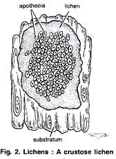
Crustose lichen క్రస్టోజ్ లైకెన్లు
Draw and write all lichens in one page
1. These lichens are found as flattened, incrustations on rocks and barks, adhering very
closely to the substratum.
2.They cannot be removed from the substratum without injuring the thallus e.g., Leconora.


1. ఇవి సూక్ష్మ పరిమాణంలో ఉంటాయి
2. ఇవి పలుచగా, పోరలుగా, బల్లపరుపుగా ఉంటాయి
3. ఆధారానికి చాలా సన్నిహితంగా అంటి పెట్టుకోని ఉంటాయి
4. కోన్ని సార్లు ఆధారంతో కలిసిపోయి ఉంటాయి


Foliose lichen పత్రాభ లైకెన్లు
1. These are flat, leaf-like, with lobed margins and are partially attached to the substratum at certain points
    like tree trunks, rocks and ground by delicate rhizines.
  2. Rhizines are hypha or hyphal tufts.


1. ఇవి పత్రాల వంటి తమ్మెలతో, ఆవాసానికి క్షితజ సమాంతరంగా పెరుగుతాయి
2. థాలస్ అధోబాగం నుంచి మూల తంతు నిర్మాణాలైన రైజిన్ లు ఏర్పడి
ఆధారంతోనికి చోచ్చుకుపోతాయి
3. థాలస్ ఉపరితలం ముడతాలు పడి ఉంటుంది.
  ఉ పార్మిలియా


Fruticose lichen  ఫ్రూటికోజ్ లైకెన్లు
1. These lichens are much branced and ribbon-like.
2. Sometimes they are filamentous and seems to be shabby so that they are named as fruticose.
3. They are filamentous, either erect e.g., cladonia or pendent e.g., Usnea
4. Fruticose lichens remain attached to the substratum only by the basal portion


1. ఇవి వస్తృతంగా శాఖీయ భవనం చెంది చిరపోదలుగా పెరుగుతాయి
2. విటి శాఖలు స్థూపాకారంలో ఆధారం నుండి పెరుగుతాయి
3. ఈ థాలస్ మూల తంతువులు ఉండవు
4. థాలస్ పీఠభాగం ఆధారానికి అంటిపెట్టుకోని ఉంటుంది.

Draw either diagram B from first picture I or
D from picture II