Note: Some of the diagrams are in either right or left side orientation, While drawing draw in correct orientation
Anomalous secondary Growth in Achyranthes Stem
The young stem has a wavy outline with alternate ridges and furrows.
Epidermis: Made up of single row of tubular cells. The cells are closely arranged with thick outer walls coated with lignin, followed by cutin. Several multicellular hairs are present over the cells of the epidermis.
Cortex: The peripheral hypodermal region is made up of collenchymas below the ridges and chlorenchyma below the furrows.
Endoermis: consists of single row of tangentially elongated parenchyma cells.
Pericycle: Made up of sclerenchyma, parenchymatous cells are also present.
Vascular Bundles: arranged in form of a ring. They are conjoint, collateral, endarch and open. Medullary rays are present between the vascular bundles.
Apart from the primary vascular bundles, two medullary vascular bundles are present in the pith region. The medullary vascular bundles are conjoint, collateral, endarch and closed. These two bundles lie and grow opposite to each other.
Secondary Growth and Medullary Bundles:
In the pericycle region, extrastelar cambium strips is seen producing secondary vascular bundles. Cambium also produces the conjunctive tissue between the vascular bundles. So phloem of the secondary vascular bundles appears in the form of patches. This phloem is the included phloem.

Anomalous secondary Growth in Boerhaavia
In the primary structure, the stem can be distinguished into epidermis, cortex and stele
Epidermis:
Single-layered epidermis consists of small, radially elongated cells. Multicellular epidermal hair arise from some cells. A thick cuticle is present on the epidermis.
Cortex:
It is well-differentiated and consists of few- layered collenchymatous hypodermis followed by chlorenchyma.Collenchyma is 3 to 4 cells deep.
Chlorenchymatous cells are thin-walled, oval, full of chloroplasts and enclose many intercellular spaces. Endodermis is clearly developed and made up of many, tubular, thick-walled cells.
Pericycle:
Inner to the endodermis is present parenchymatous pericycle but at some places it is represented by isolated patches of sclerenchyma.
Vascular Bundles:
Vascular bundles are present in three rings. In the innermost ring are present two large bundles; in the middle ring the number ranges from 6 to 14 while the outermost ring consists of 15 to 20 vascular bundles.
Vascular bundles of innermost and middle rings are medullary bundles. All the Vascular bundles are conjoint, collateral, endarch and open.
Two vascular bundles of the innermost ring are large, oval and lie opposite to each other with their xylem facing towards centre and phloem outwards.
Vascular bundles of inner and middle rings may show a little secondary growth. The cambium produces only a little amount of secondary xylem to the inner side and secondary phloem to the outer side. As a result the vascular bundles of these rings become slightly enlarged.
Anomalous Secondary Growth:
In this stem several rings of cambia are seem successively in a centrifugal manner. The abnormal cambial ring produces xylem and conjuctive tissue on the inner side and phloem and parenchyma on the outer side. The resulting tissue gives the appearance of concentric rings of vascular bundles embedded in the conjuctive tissue.

Anomalous secondary Growth in Bignonia Stem
Primary Structure:
In T.S, the young stem exhibits the ridges and furrows in outline.
Epidermis:
Single-layered epidermis consists of rectangular cells.A thick cuticle is present. A few multicellular hair are also arising from some cells.
Cortex:
It is well-differentiated into collenchyma and parenchyma. Collenchyma is present below the epidermis in the ridges in young stem but at maturity there develops sclerenchyma.
Parenchyma is present below the sclerenchyma or collenchyma in the ridges and directly below the epidermis in the grooves. Endodermis is undistinguishable from cortical cells. The cells lack casparian strips.
Pericycle:
The pericycle shows alternate bands of sclerenchyma and parenchyma.
Conjoint, collateral, endarch and open vascular bundles are arranged around the pith in form of a ring.
Anomalous Secondary Growth
At four places, the cambium produces more amount of secondary phloem on the outside and relatively small amount of secondary xylem on the inner side. As a result four deep furrows of phloem projecting into the secondary xylem are seen

Anomalous Secondary Growth in Dracaena
Primary Structure:
Epidermis: Outer most layer made of single layer of cells with thin cuticle on the outside.
Cortex: Several rows of parechymatous cortex is present.
Stele: Several vascular bundles are scattered irregulary in the ground tissue. The vascular bundles are collateral, and closed.
Anomalous Secondary growth:
During the initiation of secondary growth, the parenchymatous cells of the cortex, external to the primary vascular bundles, become meristematic and forms the cambium in a ring. The cells of cambium divide and produce more cells towards the inner side and few cells towards outside. The cells produced on the inner side develop into vascular bundles and conjuctive tissue.
Each vascular bundle develops from a single cambial initial. The initial divides first by anticlinally to form a row of two or three cells. These cells undergo periclinal division, but the division soon become irregular. This results in the formation of a group of cells.
The peripheral cells develop into xylem elements, whereas the central cells differentiate into phloem elements. This leads to formation of amphivasal vascular bundles.
Formation of cork:
After certain period of secondary growth, the parenchymatous cells below the epidermis give rise to the meristematic initials. The initials divide several times periclinally. The cells thus formed become suberised and form the cork or phellem. This cork, with suberin coated cells and radial or storied arrangement is called as storied cork.

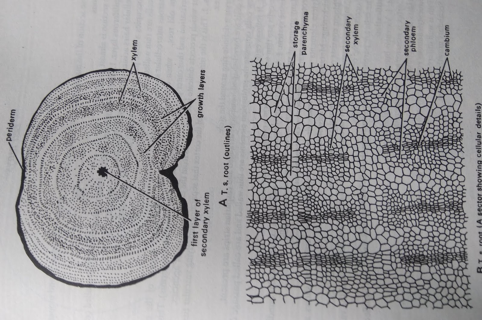
Anomalous Secondary Growth in Beta Vulgaris Root:
Periderm:
1. Outer most layer is consists of large periderm which is made up of cork (phellem), cork cambium(phellogen) and secondary cortex (phelloderm)
2. Endoermis is indistinguishable
3. Rings of vascular bundles are seen
4. Each ring has many bundles arranged very closely
5. Each vascular bundle is collateral
6. Each ring of vascular bundle is separated from the other by bands of storage parenchyma
7. Adjacent vascular bundles are also separated from each other by bands of radial parenchyma
Determination of pollen viability by Evans Blue Method
Aim :
To determine the viability of pollen grains by using Evans Blue stain method.
Principal :
Evans Blue stain penetrates into dead cell resulting in blue staining of cellular contents. The percentage of dead cells in each treatment is determined by scoring them with microscope, manually or by using a hematocytometer.
Chemicals and Requirements:
1% Evans Blue stain (dissolve one gram Evans Blue stain in hundred ml distilled water and store it in refrigerator), fresh Pollen from any plant, test tubes, centrifuge tubes
Procedure
Collect fresh pollen grains of any plant in centrifuge tube and add 1% Evans Blue stain solution to it so that it covers the pollen grains. Stir well gently with a needle and incubate for 3 minutes at room temperature. Centrifuge at 1000 RPM for 5 minutes. Discard the stain solution. Add distilled water and centrifuge as above. Repeat the washing twice or until the water is clear.
Place the pollen grains on a glass slide. Observe them under a microscope to check for the stain and unstained pollens. Count the number of stained and unstained pollens in a single view. Take readings thrice and estimate the average value of pollen grains that have retain their original colour to be reported as viable pollens. Calculate the percentage value of pollen viability. The pollen grains can be analysed with hematocytometer for systematic counting.

% of Viable Pollen grains =
No. of Viable Pollen grains. X 100
Total number of Pollen grains
% of UnViable Pollen grains =
Total number of Pollen grains
% of UnViable Pollen grains =
No. of UnViable Pollen grains. X 100
Total number of Pollen grains
Total number of Pollen grains
Isolation of Cenna Embryo
Aim: Isolation of Cenna embryo from the developing seeds.
Principal: Embryo of dicotyledons bears embryonal axis, two cotyledons, coleorhizza and coleoptiles
Apparatus:
Petridish, Blade, Slide, Forceps, Water
Procedure:
Take seeds in petridish and add some water and wait for some time. Select the developing seed and keep on the slide and remove the seed coat.
Two green colored cotyledons are seen. Place the seeds on the slide and expand the cotyledons. The embryo with two cotyledons, coleoptiles is seen.
Anomocytic Stomata:
(Irregular celled type)
1. There is no difference between the guard cells and the surrounding epidermal cells
2. Four or more cells surround the the guard cells irregularly.
3. Ex – Tridax,Amaranthus, Tagetus.

Anisocytic Stomata
( Unequal- celled type)
1. The guard cells are surrounded by 3 subsidiary cells.
2. Of the three cells, one is distinctly smaller than the other two.
3. Ex –Datura, Brassica, Hibiscus

Paracytic Stomata
( Parallel- celled type)
1. The guard cells are surrounded by two subsidiary cells.
2. The subsidiary cells are arranged parallel to the long axis of the guard cells
3. Ex – Ixora, Hamelia

Diacytic Stomata
( Corss - celled type)
1. The guard cells are surrounded by two subsidiary cells.
2. The subsidiary cells are arranged at the right angles to the long axis of the guard cells.
3. Ex – Ocimum, Hyptis, Leucas

T.S of Anther
1. It shows sporogenous tissue covered by an anther wall
2. Anther wall consists of epidermis, endothecium, middle layers and tapetum
3. Epidermis is outermost layer, one celled thick
4. Endothecium is present below the epidermis. The cells of this layer are expanded radially to form fibrous thickenings.
5. Beneath endothecium there are thin walled cells arranged in one to five layers.
6. The innermost layer of anther wall is tapetum which encircles the sporogenous tissues.
7. At four corners of the anther, four pollen chambers with sporogenous tissue are seen

Orthotropous
1. The ovule is straight, without any curvature
2. Micropyle, chalaza, funicle and embryo sac lie in a straight line.
3. Ex – Polygonum, Piper

Anatropous
1. Body of ovule becomes completely inverted to 1800.
2. The micropyle lies close to funicle
3. Micropyle, Embryo sac and chalaza lie on the same line.
4. Ex – Helianthus, Ricinus

Campylotropous
1. The body of the ovule is placed at right angles to the funiculus.
2. The body of ovule bends in such a way that micropyle comes towards funiculus.
3. Micropyle and chalaza do not lie on the same straight line.
4. Ex – Pisum, Mustard

Dicot Embryo T.S
1. The embryo consists of embryonal axis with two large lateral cotyledons.
 2. The portion of embryonal axis above the level of cotyledons is called epicotyl and the portion below the level of cotyledons is hypocotyl
3. The epicotyl forms the plumule or embryonic shoot and lower end of hypocotyl forms the radicle.
4. At one end of the embryo, swollen suspensor is seen
Monocot Embryo T.S
1. The embryo consists of shield shaped cotyledon called scutellum and the embryonal axis
2. Plumule is lateral in position and is surrounded by a leaf-sheath called coleoptile.
3. Radicle is surrounded by a root-sheath called coleorhiza

Dicot Root TS
1. It Shows Epidermis, Cortex and Stele
2. Epidermis is outermost layer made of thin walled , rectangular cells. Shows root hairs
3. Cortex is differentiated into exodermis, General cortex and Endodermis
4. Stele is smaller than cortex composed of pericycle, vascular bundles and medulla or pith
5. Radial vascular bundles are seen
6. Xylem is tetrarch and exarch.
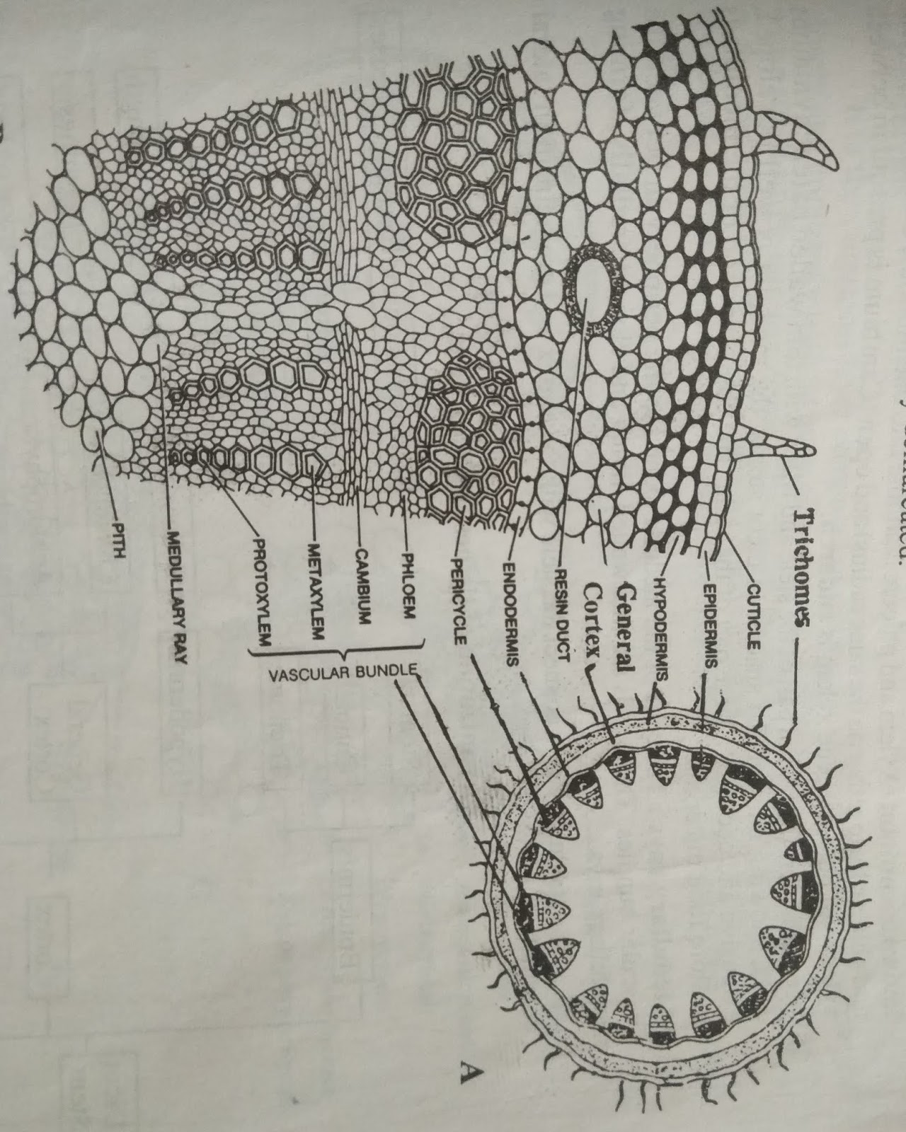
 Dicot Stem T S
1. It shows Epidermis, Cortex and Stele
2. Epidermis is outermost layer made of rectangular cells. It shows cuticle and multicellular trichomes
3. Cortex is differentiated into Collenchymatous hypodermis, parenchymatous general cortex and Enodermis
4. The stele shows Pericycle, Vascular bundles, Medulla and medullary rays
5. Wedge or top shaped vascular bundles are arranged in a ring
6. Vascular bundles are conjoint, collateral, open and endarch.

Monocot Stem T S
1. It shows Epidermis, Hypodermis, Ground tissue and Vascular bundles
2. Epidermis is outermost layer composed of rectangular cells.
3. Thick walled sclerenchymatous hyodermis is present below the epidermis
4. Below hypodermis is parenchymatous ground tissue
5. Numerous vascular bundles are scatter in the ground tissue.
6. Vascular bundles are conjoint, collateral, closed and endarch.
7. Xylem vessels are arranged in ‘Y’ Shaped and shows protoxylem lacunae

Family: Malvaceae
Pollen grains monads.
They are isopolar, radially symmetrical, spheroidal in shape.
Exine tecate, sexine thicker than nexine.
Surface beset with robust and prominent spines.

Ocimum
Family: Lamiaceae
Pollen grains are monads
They are radially symmetrical, isopolar.
Apertures - 6 colpate- Hexacolpate.
Colpi broad with blunt ends.
Examine sub - tectate, surface prominently reticulated.

Acacia
Family: Mimosoidae
Pollen grains monads
Radially symmetrical arranged in polyads of 16 - grains.
Each grain spheroidal to sub- spheroidal
Each grain provided with 2 or 3 faint pores often considered as inaperturate.
Exine tecate, sexine thicker than nexine
Surface granular

Grass Pollen grains
Family: Poaceae
Pollen grains monads
Radially symmetrical, heterpolar, spheroidal.
Distally monoporate; pore with a prominent annulus
Exine tectate, Sexine as thick or thicker than nexine.
Exine pilate (smooth) to faintly granular

(Irregular celled type)
1. There is no difference between the guard cells and the surrounding epidermal cells
2. Four or more cells surround the the guard cells irregularly.
3. Ex – Tridax,Amaranthus, Tagetus.

Anisocytic Stomata
( Unequal- celled type)
1. The guard cells are surrounded by 3 subsidiary cells.
2. Of the three cells, one is distinctly smaller than the other two.
3. Ex –Datura, Brassica, Hibiscus

Paracytic Stomata
( Parallel- celled type)
1. The guard cells are surrounded by two subsidiary cells.
2. The subsidiary cells are arranged parallel to the long axis of the guard cells
3. Ex – Ixora, Hamelia

Diacytic Stomata
( Corss - celled type)
1. The guard cells are surrounded by two subsidiary cells.
2. The subsidiary cells are arranged at the right angles to the long axis of the guard cells.
3. Ex – Ocimum, Hyptis, Leucas

T.S of Anther
1. It shows sporogenous tissue covered by an anther wall
2. Anther wall consists of epidermis, endothecium, middle layers and tapetum
3. Epidermis is outermost layer, one celled thick
4. Endothecium is present below the epidermis. The cells of this layer are expanded radially to form fibrous thickenings.
5. Beneath endothecium there are thin walled cells arranged in one to five layers.
6. The innermost layer of anther wall is tapetum which encircles the sporogenous tissues.
7. At four corners of the anther, four pollen chambers with sporogenous tissue are seen

Orthotropous
1. The ovule is straight, without any curvature
2. Micropyle, chalaza, funicle and embryo sac lie in a straight line.
3. Ex – Polygonum, Piper

Anatropous
1. Body of ovule becomes completely inverted to 1800.
2. The micropyle lies close to funicle
3. Micropyle, Embryo sac and chalaza lie on the same line.
4. Ex – Helianthus, Ricinus

Campylotropous
1. The body of the ovule is placed at right angles to the funiculus.
2. The body of ovule bends in such a way that micropyle comes towards funiculus.
3. Micropyle and chalaza do not lie on the same straight line.
4. Ex – Pisum, Mustard

Dicot Embryo T.S
1. The embryo consists of embryonal axis with two large lateral cotyledons.

3. The epicotyl forms the plumule or embryonic shoot and lower end of hypocotyl forms the radicle.
4. At one end of the embryo, swollen suspensor is seen
Monocot Embryo T.S
1. The embryo consists of shield shaped cotyledon called scutellum and the embryonal axis
2. Plumule is lateral in position and is surrounded by a leaf-sheath called coleoptile.
3. Radicle is surrounded by a root-sheath called coleorhiza

Dicot Root TS
1. It Shows Epidermis, Cortex and Stele
2. Epidermis is outermost layer made of thin walled , rectangular cells. Shows root hairs
3. Cortex is differentiated into exodermis, General cortex and Endodermis
4. Stele is smaller than cortex composed of pericycle, vascular bundles and medulla or pith
5. Radial vascular bundles are seen
6. Xylem is tetrarch and exarch.

1. It shows Epidermis, Cortex and Stele
2. Epidermis is outermost layer made of rectangular cells. It shows cuticle and multicellular trichomes
3. Cortex is differentiated into Collenchymatous hypodermis, parenchymatous general cortex and Enodermis
4. The stele shows Pericycle, Vascular bundles, Medulla and medullary rays
5. Wedge or top shaped vascular bundles are arranged in a ring
6. Vascular bundles are conjoint, collateral, open and endarch.
1. It shows Epidermis, Hypodermis, Ground tissue and Vascular bundles
2. Epidermis is outermost layer composed of rectangular cells.
3. Thick walled sclerenchymatous hyodermis is present below the epidermis
4. Below hypodermis is parenchymatous ground tissue
5. Numerous vascular bundles are scatter in the ground tissue.
6. Vascular bundles are conjoint, collateral, closed and endarch.
7. Xylem vessels are arranged in ‘Y’ Shaped and shows protoxylem lacunae

Palynology
HibiscusFamily: Malvaceae
Pollen grains monads.
They are isopolar, radially symmetrical, spheroidal in shape.
Exine tecate, sexine thicker than nexine.
Surface beset with robust and prominent spines.

Ocimum
Family: Lamiaceae
Pollen grains are monads
They are radially symmetrical, isopolar.
Apertures - 6 colpate- Hexacolpate.
Colpi broad with blunt ends.
Examine sub - tectate, surface prominently reticulated.

Acacia
Family: Mimosoidae
Pollen grains monads
Radially symmetrical arranged in polyads of 16 - grains.
Each grain spheroidal to sub- spheroidal
Each grain provided with 2 or 3 faint pores often considered as inaperturate.
Exine tecate, sexine thicker than nexine
Surface granular

Grass Pollen grains
Family: Poaceae
Pollen grains monads
Radially symmetrical, heterpolar, spheroidal.
Distally monoporate; pore with a prominent annulus
Exine tectate, Sexine as thick or thicker than nexine.
Exine pilate (smooth) to faintly granular

Tangential Longitudinal
Section (TLS)
1. In this section, the cells of the axial system are cut longitudinally and those of the ray system at right angles to their orientation.
2. The width and height of the ray are clearly visible
3.
Radial Longitudinal Section (RLS)
Marvel
ReplyDeleteNice
ReplyDeleteHmmmm
ReplyDeleteMantram Nursing Academy is a leading BSc Nursing coaching center in Patiala. With experienced teachers and modern teaching methods, they prepare students for competitive exams through interactive sessions and rigorous practice.
ReplyDeleteBSc Nursing Coaching Center in Patiala